Mohs Micrographic Surgery & Reconstruction
Mohs micrographic surgery is the gold standard for treatment of basal cell and squamous cell carcinomas, providing patients with medical, cosmetic and financial advantages. Unlike traditional skin cancer surgery, which only evaluates only about 1% to 3% of the skin cancer margins, the Mohs technique evaluates the complete surface area for malignancy. Mohs surgery has an unprecedented cure rate of up to 99% for skin cancer. This highly specialized and precise surgical technique results in the most healthy surrounding skin being spared and the smallest wound possible.
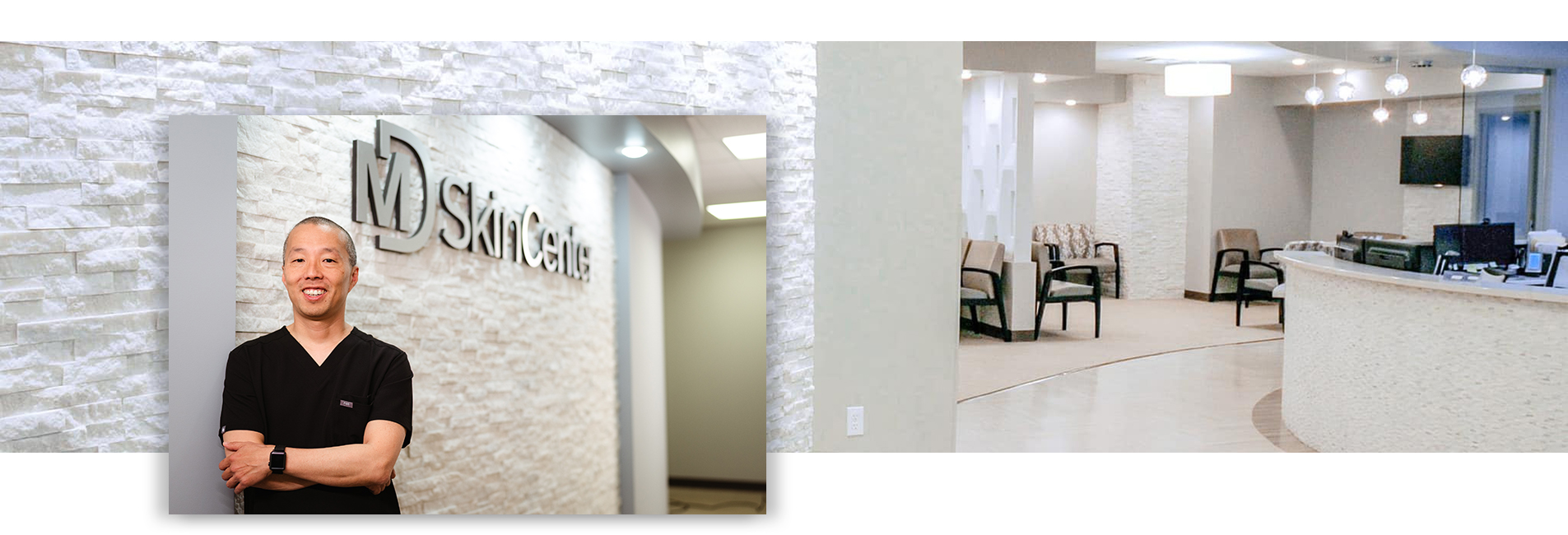
Depending on the extent of the cancer, most wounds require some surgical repair after Mohs surgery. As a board certified head and neck facial plastic surgeon, Dr. Andrew Jun is unlike most Mohs surgeons in that he is not only capable of removing the skin cancer but also performing customized reconstructions. His high level of training and surgical skills provide patients with the most optimal cosmetic and functional outcomes. Over the past 20 years, Dr. Jun has dedicated his career to removing and reconstructing tens of thousands of skin cancers. Dr. Jun and the MD SkinCenter team understand how a diagnosis of skin cancer can be frightening and overwhelming for patients. Their goal is to provide the highest level of care in the treatment of your skin cancer with the warmth and compassion you would expect from your provider.
Mohs surgery is a highly specialized treatment for complex skin cancers performed by Dr. Jun. Distinct from surgical excision, the Mohs technique involves removing layers of tissue. The tissue is then mapped, color-coded and processed onto microscope slides for examination. On the same day of surgery, Dr. Jun will evaluate 100% of the tissue margin to ensure that the entire skin cancer is removed. Additional layers are taken, as needed, until all of the tumor is completely removed.
The majority of tumors treated with Mohs surgery are complex basal cell carcinomas and squamous cell carcinomas. Other uses include more rare skin cancers, as well as thin melanomas. Skin cancers may be considered complex when:
- the cancer is in an area where preservation of healthy tissue is critical to minimize scarring and maximize function and cosmesis
- the cancer is in an area of higher tumor recurrence (ears, lips, nose, eyelids, temples)
- the cancer is in a previously treated area and is recurrent
- the cancer is very large
- the margins of the cancer are not well defined
- the patient is immunosuppressed (e.g. organ transplantation, HIV infection, chronic lymphocytic leukemia)
Unlike other methods of treatment, Mohs surgery is widely regarded as the most successful and effective method of treating skin cancer with the highest cure rate of up to 99%. The tissue that is removed is microscopically examined to confirm complete tumor removal. If microscopic examination reveals additional cancer cells, it will show the surgeon the precise location where additional removal is needed.
The procedure maximizes both functional and cosmetic outcomes since it results in the smallest wound and spares the most healthy skin and tissue surrounding the tumor. When Dr. Jun performs the reconstruction after the Mohs surgery, he also creates a reconstructive surgical plan tailored to the patient, wound size and location, to provide minimal scarring and maximum function.
Performed in an outpatient setting under local numbing, patients avoid the time, expense and side effects of inpatient surgery and general anesthesia. Patients also appreciate knowing the results the same day and having peace of mind that the cancer is completely removed.
Step 1: Anesthesia: the tumor site is numbed using local anesthesia. General anesthesia is not necessary for Mohs surgery.
Step 2: Removal of visible tumor is then performed by surgically removing a thin, saucer shaped “layer” of tissue.
Step 3: Mapping the tumor using orientation to landmarks (eye, nose, etc) and color-coding each section to match.
Step 4: Evaluation of processed tissue by the Mohs surgeon who will determine if margins are clear, or alternatively if additional “layers” will need to be removed to clear the tumor.
Step 5: Additional stages taken if needed to ensure all cancer cells are removed.
Step 6: Reconstruction of the wound created by removal of the tumor. The best method of repair is chosen only after the cancer has been completely removed. Stitches may be used to close the wound side-to-side, or a graft or flap may be designed. In some instances, a wound may heal very well naturally.
